In order to combat the effect of any new virus, it is important to understand the mechanism by which it infects host cells, overcomes the host’s immune defense mechanism, and improves viral protein synthesis and replication.
To study: Key features of the SARS-CoV-2 and NSP1 leader required for viral escape from NSP1-mediated suppression. Image Credit: Design_cells / Shutterstock.com
Background
The most important preventive approach to minimize the effect of Severe Acute Respiratory Syndrome Coronavirus 2 (SARS-CoV-2) infection in host cells has so far been the development of vaccines. These vaccines target the viral spike protein responsible for binding to host cells and entering the systemic circulation.
However, vaccination does not completely stop the spread of the disease. Because SARS-CoV-2 is a new virus, it is inevitable that it undergoes several sets of mutations that produce new variants that may be resistant to existing vaccines.
This highlights the need to develop drugs targeting the main machinery of SARS-CoV-2 that can be used to treat infected people. To this end, non-structural protein 1 (NSP1) appears to be an ideal candidate as a target. Previous research has shown that NSP1 is a conserved region in all beta-coronaviruses, the family to which SARS-CoV-2 belongs.
However, previous reports in the literature on the methods by which NSP-1 invades the host system stops the synthesis of host-specific proteins and induces the synthesis of viral proteins remains contradictory. In a recent study published on the Preprint Server, bioRxiv*, researchers are attempting to resolve this confusion by studying the molecular aspects of entry into the host cell and inhibition of host-specific cellular mechanisms.
What is NSP1?
NSP1 is an important virulence factor that plays a crucial role in the pathogenicity of SARS-CoV-2 by helping the virus evade the host’s innate immune response. NSP1 downregulates the expression of host genes and promotes its own spread by neutralizing the antiviral responses of the host cells to which the virus binds.
The SARS-CoV-2 genome is a positive-stranded ribonucleic acid (RNA) virus of about 30 kilobases (kb) with a 5′-cap, 5’UTR (or leader), 3’UTR and a structure. polyA tail. SARS-CoV-2 contains 10 open reading frames (ORFs) encoding proteins, which are parts of the genome where protein translation can occur without encountering stop codons.
Upon entry into the cell, genomic RNA (gRNA) is translated into polyprotein, which is transformed into 16 non-structural proteins (NSPs). Subsequently, its gRNA serves as a template to generate a set of subgenomic mRNAs (sgRNAs) that encode other viral proteins.
The first protein produced by coronaviruses upon infection is NSP1, which is encoded by ORF1a at the 5 ‘end of the gRNA. In previous studies on related SARS-CoV-1, NSP1 was found to inhibit host immune responses by repressing host transcript expression and preventing complete induction of interferon ( IFN) and STAT1 phosphorylation, both of which are crucial for the initiation of antiviral immune responses.
About the study
In the present study, researchers assessed the potential of NSP1 as a target by studying the detailed molecular mechanisms of its functions. To this end, they synthesized the viral genome by cloning known sequences of SARS-CoV-1 and 2 and separating specific proteins using Western Blot techniques.
Researchers used mutagenesis to detect the effect of different mutations in specific genes to assess their roles in immune evasion by SARS-CoV-2 NSP1. Reporter assays have also been used to detect specific changes resulting from induced mutations. Mass spectroscopy was used to analyze proteins.
Specific biotin-based assays (BioID) were used to locate the proteins of interest and study the different proteins in the functional parts of the SARS-CoV-2 NSP1 protein. This test helped researchers understand the different parts of the NSP1 protein involved in different antiviral defense mechanisms. For all of these experiments, the researchers used human HEK293T cells as hosts.
Study results
Researchers in the present study found that stem loop-1 (SL1), which is a hairpin structure in single-stranded RNA that helps viral mRNA maintain its structure and pursue specific enzymatic interactions , is both necessary and sufficient to escape the mediation of NSP1. repression. Mutagenesis was used to isolate 3 proteins specific for cytosine, which is one of the four nucleic acid bases of RNA, which were crucial for evading host defense mechanisms.
After further analysis of the protein by BioID, it was discovered that NSP1 blocked host translation by inserting its C-terminal domain into the mRNA entry tunnel on the 40S ribosomal subunit. In doing so, NSP1 blocked the ribosomal synthesis of the host protein and exported viral mRNAs into the system. An artificially constructed non-functional mutant KH164AANSP1 was unable to mimic the mechanism of wild-type NSP1 as efficiently.
Normally, when a virus attacks host cells, viral RNA is detected by host RNA helicases in infected cells, resulting in activation of the transcription factors ATF2 / c-Jun, IRF3 / IRF7 and NF-. kB. These factors then induce the production of cytokines, including members of the IFN family, which bind to their respective receptors and trigger a second wave of cytokine signaling. These two waves upregulate genes that inhibit viral replication.
Ultimately, NSP-1 was found to escape these pathways to prevent cytokine induction, thereby helping the virus to spread through the host system.
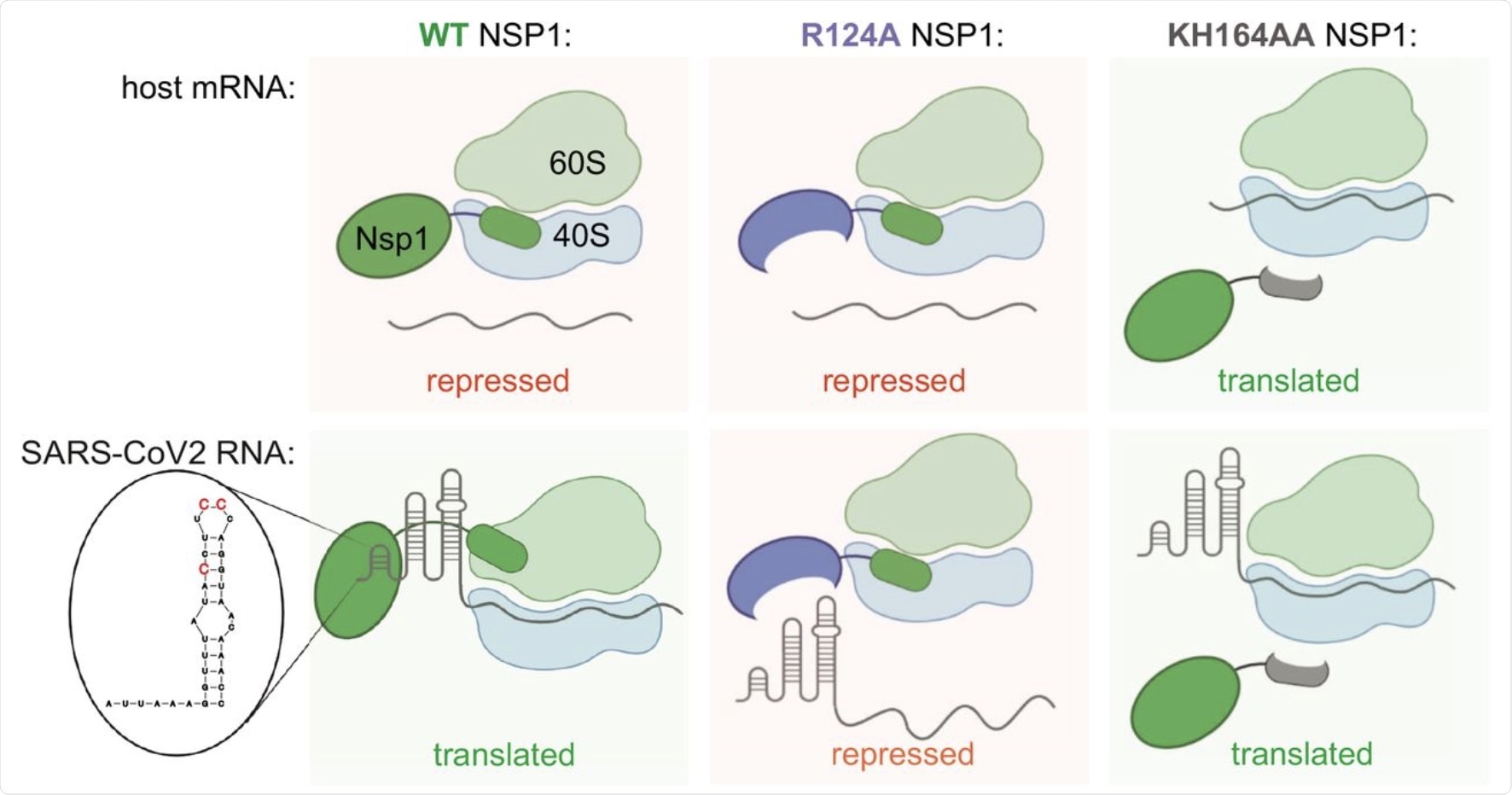 Speculative model for NSP1 function in host mRNA repression and activation of SARS-CoV-2 expression. The WT NSP1 protein represses host mRNA translation by blocking the ribosome entry tunnel. SARS-CoV-2 genomic and subgenomic RNAs escape repression by NSP1 due to SL1 in their 5’UTR, interacting with NSP1. In this way, viral RNA hijackings harbor ribosomes without competing to limit eIF in infected cells. In particular, the positions C15, C19 and C20 are necessary to attenuate the silence NSP1. The K164H165 positions in NSP1 are required for interaction with the ribosome, therefore the KH164AA mutant of NSP1 does not repress the host or SARS-CoV-2 mRNA. The R124 position is required for interaction with SL1, therefore the NSP1 R124A mutant represses both host and viral mRNA.
Implications
The present study showed that wild-type SARS-CoV NSP1 was responsible for binding to host ribosomes and inducing viral mRNA production. NSP1 has also been shown to evade immune responses and delay the release of inflammatory cytokines.
The first therapeutic target identified in this study is the NSP1 binding site to the host ribosome,. A second target would consist of inducing mutations in the NSP1 region which interacts with the viral leader to escape the host’s antiviral responses, and that in the SL-1 region, by targeting the 3 specific cytosine molecules.
*Important Notice
bioRxiv publishes preliminary scientific reports which are not peer reviewed and, therefore, should not be considered conclusive, guide clinical practice / health-related behavior, or treated as established information